Go to last entry.......................................Go to previous entry
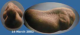
14 March - Cold weather has meant that for the last two days I have not seen a single frog. On the positive side, the pond has been topped up with rain water, and with it a useful input of oxygen for the developing frog spawn. The embryo is becoming more elongated and the myotomes are a bit clearer, but I'm not yet sure about the structures forming at the head end.
With the temperature creeping back up (9C at 4.15pm) a couple of frogs have ventured to the water's surface, and I could hear one croaking.
16 March - Today's embryo picture shows more detailing on the surface of it. The blurred green spots are algae growing on the glass of the tank. I did not spend much time looking at the pond today, but there were a few frogs about when I made a quick check in the morning.
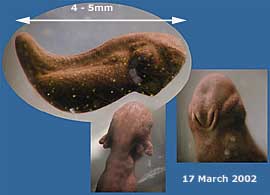
The picture shows how the external gills are starting to develop and the right hand image shows what looks like a mouth. This is, in fact the Mucus Gland which the larva uses to attach itself to weeds after it has hatched and is free of the jelly. It does not yet have a mouth opening. In the pond many of the larvae are already free of the jelly and are starting to mass in dark clumps. If you click on the image you can see a larger version.
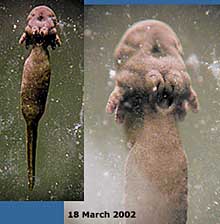
18 March - I left the taking of this picture until late this evening as a result of my pre-occupation with my sick laptop. I cannot tell which is the larva that I've been following. There are a group of them attached to the glass by their Mucus glands. The 'dimple' above the gland is where the mouth will open up after about a week. In the meantime, the tadpole will continue to feed on its yolk.
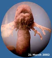
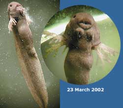
21 March - After a gap of a couple of days the young tadpoles are now all free of their jelly and are mostly attached to the glass. They occasionally wriggle about to adjust their positions. The picture of a typical one shows how its mouth is now open, although not functioning yet and its external gills have become almost antler-like. The Mucus glands are less prominent and at the side of the head you may see where an eye is developing. Click on the picture to see a larger version.
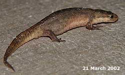
This newt nearly got itself trodden on tonight. It was on the decking just outside our kitchen door. I moved it back to the shelter of the plants near the pond. In the pond, while the few frogs I can see seem to be in a state of well earned rest, the newts are as active as ever under the shelter of darkness. However, I can see no signs of egg-laying as yet.
The mouth continues to develop and you can see its horny jaws forming. these will bear horny teeth, used as rasps to scrape off mainly vegetable matter. You can also see how the tail has become the familiar tadpole shape. Click on the picture to see a larger version. I have now split the March diary into two parts to speed up downloading.
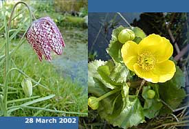
Click on the picture to see a larger version. Last year, my first sighting of newt egg laying was on 1 April.
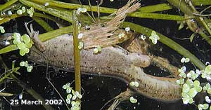
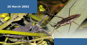
Invertebrate life in the pond is really getting going now. As I watched the newts I saw five water scorpions creeping about in the weeds, and my first pond skater of the year. Better picture opportunities will come in the weeks ahead.
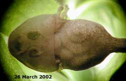
You can now see the coiled gut. The external gills can be seen protruding from a fold of skin which runs right under the tadpole. This fold is becoming a gill chamber. The mucus gland (behind the mouth) is much less evident now.
Click on the picture to see a larger version.
The small object you can see about half way back along the main part of its body is called the Branchial opening, or spiracle. It is only found on the left side of the tadpole and is an opening by which its gill chamber opens to the outside, the external gills being replaced by internal ones. Click on the picture to see a larger version. |
|
 15
March - I had problems
trying to get a clear image of the embryo today, so this picture
is of its 'next door neighbour', which looked very similar.
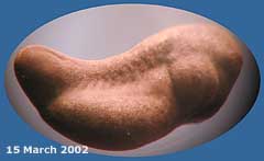
15
March - I had problems
trying to get a clear image of the embryo today, so this picture
is of its 'next door neighbour', which looked very similar.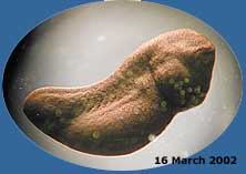
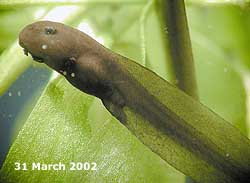
 Taking
close-up pictures of the tadpoles is now a bit of a challenge
as they do not stay still for very long. This one swam off moments
after the shutter was pressed. They are now about 1cm long.
Taking
close-up pictures of the tadpoles is now a bit of a challenge
as they do not stay still for very long. This one swam off moments
after the shutter was pressed. They are now about 1cm long.